


This disease is misunderstood, although thousands of observations have been described with diagnosis and subsequent treatment.
The diversity and non-specificity of the clinical picture of small pelvic varicose veins leads to gross errors on the part of the diagnosis, which will influence the consequences in the future.
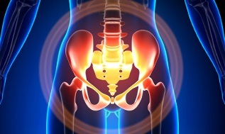
Characteristics of varicose veins in small pools
Pelvic veins are several times longer than arteries, leading to greater capacity. This is due to the phylogeny of the vascular system of the pelvic region. The pelvic veins are highly adaptive and prone to transformation, which contributes to the formation of a densely intertwined network.
The speed and direction of blood flow is regulated by valves controlled by complex humoral mechanisms. The valves equalize the pressure in different parts of the venous network.
When the valves do not perform their functions, blood stagnation develops, leading to vascular pathology and varicose veins. The uniqueness of the pelvic veins lies in the fact that the wide ligaments of the uterus, which hold the lumen of the vessel wide, can narrow, causing pathology.
Causes of Occurrence
The cause of abnormal venous dilatation in the pelvis is as follows:
- Disruption of blood flow;
- Abolition of the vein trunk;
- Compression of the parotid wings with a changed position of the uterus, such as during post-reflection;
- ovarian venous valve insufficiency (congenital or acquired);
- Obstructive postphlebitic syndrome;
- Connective tissue pathology;
- Arteriovenous angiodysplasia;
- Prolonged sitting, hard physical work;
- Varicose veins in lower limbs;
- Pregnancy (3 or more) and childbirth (2 or more);
- Diseases of the female genital organs (chronic salpingo-oophoritis, ovarian tumors, uterine fibroids and genital endometriosis);
- Adhesion of pelvic organs;
- Obesity.
Classification by disease severity
Based on the size of the dilated vein, the following degrees are distinguished:
- not more than 0, 5 cm, "corkscrew" vessel flow;
- 0, 6-1 cm;
- more than 1 cm.
Variations in the course of the disease
- varicose veins of the perineum and vagina;
- syndrome in pelvic venous congestion;
Symptoms
- The most common - frequent pains in the lower abdomen, perineum after long static and dynamic overload. The pain increases in the second stage of the cycle, after hypothermia, fatigue, stress, exacerbation of various diseases.
- Feels "wrong", pain during and after sex.
- Dysmenorrhea - menstrual disorders, including pain.
- Glands of the genital tract to a greater extent than normal.
- Stagnation of blood leads to infertility, miscarriage, abortion.
- Violation of urination due to dilation of venous veins.
Diagnostics
Diagnosis of the disease with complaints only is successful in only 10% of cases.
Touching the inside walls of the pool allows you to feel elongated seals and venous nodules. Seen from the mirrors, cyanosis of the vaginal mucosa can be seen.
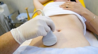
The method of choice is an ultrasound scan with color Doppler imaging, which allows the detection of not only varicose ovarian veins but also venous thrombosis, post-thrombophlebitic obstructions. Ultrasound shows tortuity, "worm-like" structures, without signal reflection, localized on the lateral surface of the uterus.
The Doppler effect is based on the "shade" of blue and red, venous and arterial blood flow.
The ultrasound device uses a special program to detect the movement of blood from the sensor and in the other direction, calculate the speed of blood flow and the type of vessel.
But the exact definition of the vein or artery remains with the doctor. The Doppler method works in almost all cases, exceptions to the rules are determined by our body, because the blood flowing from the heart is not always arterial and vice versa.
Thus, an ultrasound diagnostician sees this arterial or venous vessel, its size, blood flow rate, and many indicators that an ordinary person does not need but plays an important role in making a diagnosis. Transabdominal and transvaginal sensors are used for this.
In 5. 7% of cases, the disease is accidentally detected during screening. Usually the ovary vein has a diameter of 0, 4 cm.
CT and MRI are very accurate. These methods make it possible to detect the accumulation of varicose veins in the ligaments around the uterus, ovaries, and these organs. It is possible to determine concomitant pathology.
Phlebographic research is a very reliable method.
Contrast is performed at the height of the Valsalva test against blood flow. This allows you to see the valve failure accurately.
Left rethngenorenoscopy, renal phlebography, superselective phleboovarioscopy, and phleboovariography are also used on both sides. These methods make it possible to determine hemodynamic and anatomical changes in venous veins and at the sites where sex veins flow.
Superselective fleboovarioscopy is performed by catheterization of the genital veins through the contralateral femoral or subclavian vein, followed by contrast injection.
Blood from the varicose veins of the uvine plexus is thrown through the ovarian vein. But in case of high blood pressure, it enters the inner hip vein through the extraorganic uterine veins. The plexus of the veins through which outflow can occur includes the sacral and bladder plexus.
Left phleboovaricography has 3 stages of venous stasis in the uviform plexus of the left ovary:
- There is no outflow from the left ovarian plexus or an additional short path follows.
- There is another long way to go.
- Two additional outflow paths are shown, or one additional and additional.
In stages 2 and 3, varicose veins of the uviform plexus of the right ovary develop.
Laparoscopy is used for differential diagnosis. The abnormally tortuous veins are located in the region of the ovary, in the direction of the round and broad ligaments. They look like a large thin and tense-walled cyanotic conglomerates.
The complexity of the diagnosis lies in the fact that the disease often hides behind the signs of an inflammatory process, differs in its clinical manifestations, disguises itself as endometriosis, visceral prolapse, postoperative neuropathies and many extragenital diseases.
Management
The main goal of treatment is to remove reflux in the veins. Conservative treatment is used in the early stages of the disease. In the later stages of the disease, surgery is the treatment of choice.
Conservative treatment

It consists of normalizing venous tone, improving hemodynamics and trophic processes.
Treat symptomatically with individual symptoms. Non-steroidal anti-inflammatory pain, bleeding - hemostatic therapy.
The main drugs for conservative treatment are venotonic drugs and platelet inhibitors.
Phlebotonics - improves the sound of the vessel wall and increases blood flow. For this disease, it is better to consult a gynecologist about certain medications.
Physiotherapy is an important method.
Surgical treatment
- Resection of the varicose vein.
- Gonado-caval reversal.
- Sclerosis in laparoscopy.
- Ovarian venous occlusion by X-ray endovascular methods.
Folk remedies
Because the weakness of the valve device is a major factor in the development of the disease, all folk remedies applied to the varicose veins of the lower extremities are also used.
Commonly used are hazelnuts, hops, nettles, horse chestnuts, dandelion root, kombucha, willow, oak, St. John's wort, string, pollen and many other plants.
Effective: treatment with baths oak, chestnut, willow, chamomile, pharmacy, dried herbs, St. John's wort, string.
Prevention
- If you have any of the complaints, predictions, or illnesses listed above, contact your gynecologist first.
- It is necessary to normalize the work schedule and rest, try not to stay in a vertical position for a long time, physical overload.
- Practice prevention exercises: "pedal", "birch", "scissor legs"
- Follow your diet: eat foods high in vitamins E, P, C, try to eat only white meat, less fatty, replace it with fruits, vegetables, cereals.
- Drink plenty of fluids, but at least 1. 5 liters per day.
- Get rid of excess weight, bad habits.
- Talk to your doctor about wearing compression garments, this will improve blood flow from your lower extremities, thus reducing pelvic congestion.
- Avoid baths, saunas, steam baths, hot tubs.
In order to avoid becoming ill with such a difficult-to-diagnose disease, you should follow the preventive recommendations listed above. Treat your health as the most valuable thing in life.
For the slightest suspicious symptoms that you cannot get rid of within a few days, see your doctor. He must provide you with highly trained help and save you from suffering.